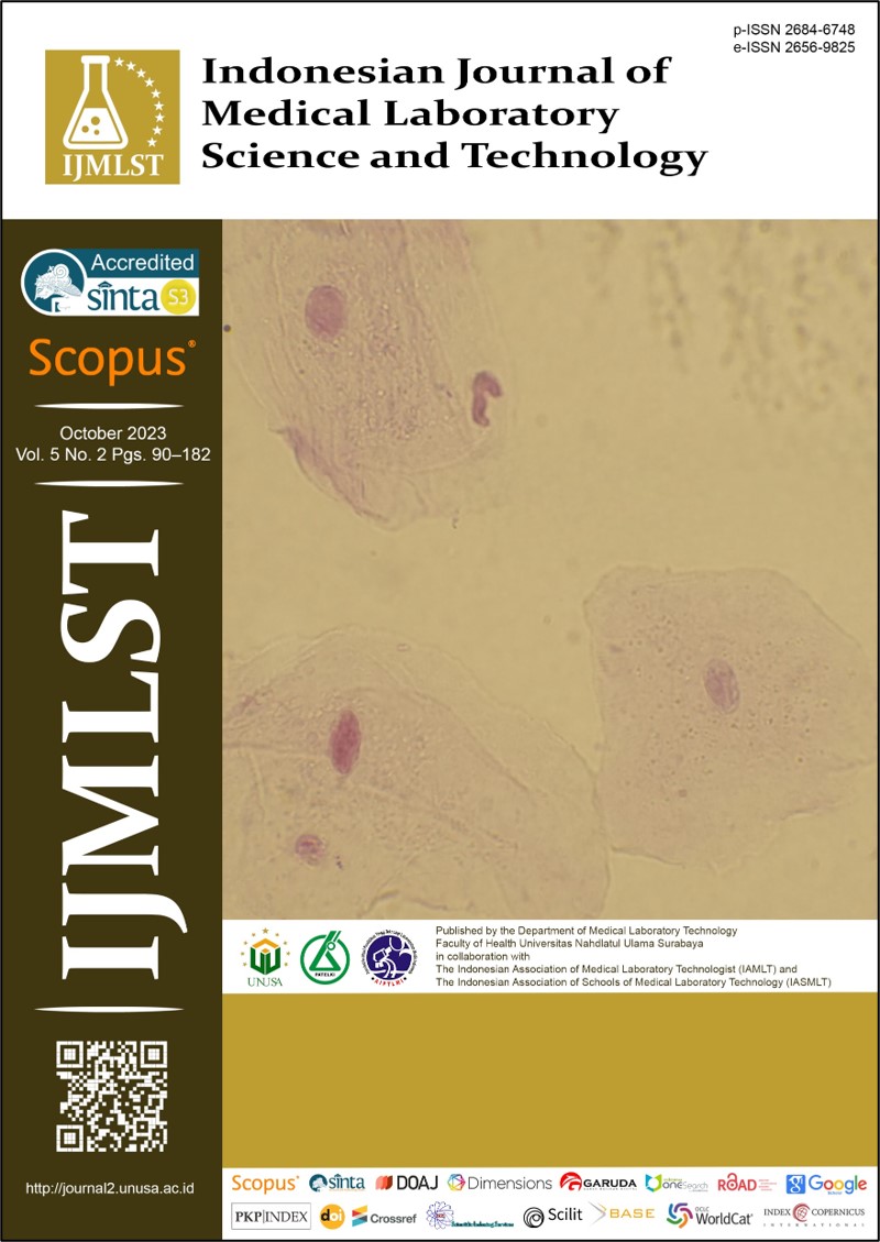
Histopathological of Mice (Mus musculus) Liver Induced by Lead (Pb) Orally
Main Article Content
Abstract
Lead (Pb) is a prominent heavy metal emitted by motor vehicle exhausts, factory and mining fumes. Its presence in the atmoshpere can endure for up to seven days, posing a considerable risk of contaminating surrounding food and beverages. Lead enters the body through inhalation and the skin. Lead can also enter the human body via the oral route and accumulate in the body. It causes health problems such as oxidative stress and damage human organs such as the kidneys and liver. This research aims to examine the effect of oral lead exposure on the liver histopathology of Swiss Webster strain mice (Mus musculus). Employing a non-probability sampling technique, 25 male mice were divided into 5 groups: negative control, K2, K3, K4 and K5. These mice were administered a daily oral dose of 0.5 mL and subsequently euthanized in CO2 chamber the following week for liver dissection. The findings reveal signs of hydropic degeneration characterized by cellular swelling, irregular shapes, and disrupted organelles in groups K2, K3, K4, and K5. In addition, the mean degree of liver damage was observed as 0 for the negative control, 1 for group K2, 1 for group K3, 2 for group K4, and 3 for group K5. In conclsuin, this study confirms that lead exposure can result in dentrimenal liver histopathology changes in mice.
Downloads
Article Details
Copyright (c) 2023 Liah Kodariah, Pakpahan Suyarta Efrida, Nugraha Aditya, Zalzabila Rena Nurzal

This work is licensed under a Creative Commons Attribution-ShareAlike 4.0 International License.
References
Kordas K, Ravenscroft J, Cao Y, McLean EV. Lead exposure in low and middle-income countries: Perspectives and lessons on patterns, injustices, economics, and politics. Int J Environ Res Public Heal. 2018;15(11):2351. DOI: 10.3390/ijerph15112351 DOI: https://doi.org/10.3390/ijerph15112351
Greenstone M, Fan Q. Indonesia’s worsening air quality and its impact on life expectancy. Air Qual Life Index. 2019:1–10.
Rinawati D, Barlian B, Tsamara G. Identification of lead (Pb) levels in the blood of gas station operator officers 34-42115 Serang City. [Identifikasi kadar timbal (Pb) dalam darah pada petugas operator SPBU 34-42115 Kota Serang]. J Med. 2020;7(1):1–8. DOI: 10.36743/medikes.v7i1.195 DOI: https://doi.org/10.36743/medikes.v7i1.195
Manisalidis I, Stavropoulou E, Stavropoulos A, Bezirtzoglou E. Environmental and health impacts of air pollution: A Review. Front Public Heal. 2020;8:14. DOI: 10.3389/fpubh.2020.00014 DOI: https://doi.org/10.3389/fpubh.2020.00014
Ogun AS, Joy NV, Valentine M. Biochemistry, heme synthesis. StatPearls. 2023; Available from: https://www.ncbi.nlm.nih.gov/books/NBK537329/
Rosita B, Program L, Analis S, Stikes K. Padang p. relationship between lead (Pb) toxicity in the blood and hemoglobin of Pekanbaru motorbike painting workers. [Hubungan toksisitas timbal (Pb) dalam darah dengan hemoglobin pekerja pengecatan motor pekanbaru]. Pros Semin Kesehat PERINTIS. 2018;1(1):2622–2256. Available from: https://jurnal.upertis.ac.id/index.php/PSKP/article/view/64
Hopkins J. Liver: Anatomy and functions | Johns Hopkins Medicine [Internet]. 2023. Available from: https://www.hopkinsmedicine.org/health/conditions-and-diseases/liver-anatomy-and-functions
Rachmani SD, Safitri E, Darsono R, Hestianah EP, Plumeriastuti H, Santoso KP. The effect of administering bee propolis on the histopathological appearance of the liver of female mice (Mus musculus) exposed to the heavy metal Pb acetate [Pb(C2H3O2)2]. [Pengaruh pemberian propolis lebah terhadap gambaran histopatologi hepar mencit (Mus musculus) betina yang dipapar logam berat Pb asetat [Pb(C2H3O2)2]]. J Basic Med Vet. 2019;8(1):21–8. DOI: 0.20473/.v8i1.19988 DOI: https://doi.org/10.20473/.v8i1.19988
Rosmaidar R, Nazaruddin N, Winaruddin W, Balqis U, Aransyah TA. The effect of lead (Pb) exposure to the histopathology of nile tilapia (Oreochromis nilloticus) liver. [Pengaruh paparan timbal (Pb) terhadap histopatologis hati ikan nila (Oreochromis nilloticus)]. J Ilm Mhs Vet. 2017;1(4):742–8. DOI: 10.21157/jim%20vet..v1i4.4992
Jarrar BM, Taib NT. Histological and histochemical alterations in the liver induced by lead chronic toxicity. Saudi J Biol Sci. 2012;19(2):203–10. DOI: 10.1016/j.sjbs.2011.12.005 DOI: https://doi.org/10.1016/j.sjbs.2011.12.005
Hidayati DMN, Athiroh N. P Histopathologic profile in the heart tissue of 90-days sub-chronic rats using metanolic extract of Scurrula atropurpurea (Bl.) Dans. BIOSAINTROPIS. 2017;3:30–6. DOI: 10.33474/e-jbst.v3i2.127
Grada A, Mervis J, Falanga V. Research techniques made simple: Animal models of wound healing. J Invest Dermatol. 2018;138(10):2095-2105.e1. DOI: 10.1016/j.jid.2018.08.005 DOI: https://doi.org/10.1016/j.jid.2018.08.005
Pusparani G, Desnita E, Edrizal E. Effect of red andong leaf extract Cordyline fruticosa (L) A. chev on the speed of topical wound closure in white mice (Mus musculus). [Pengaruh ekstrak daun andong merah Cordyline fruticosa (L) A. chev terhadap kecepatan penutupan luka secara topikal pada mencit putih (Mus musculus)]. B-Dent, J Kedokt Gigi Univ Baiturrahmah. 2018;3(1):59–67. DOI: 10.33854/JBDjbd.39 DOI: https://doi.org/10.33854/JBDjbd.39
Kodariah L, Wahid AA. Effect of coriander seed extract (Coriandrum sativum) on triglyceride levels and liver histology of rats (Rattus novergicus) induced by high-fat diet. [Pengaruh ekstrak biji ketumbar (Coriandrum sativum) terhadap kadar trigliserida dan gambaran histologi hati tikus (Rattus novergicus) yang diinduksi oleh pakan tinggi lemak]. J Biotek Medisiana Indones. 2020;9(1):47–54. DOI: 10.22435/jbmi.v9i1.3899 DOI: https://doi.org/10.22435/jbmi.v9i1.3899
Kodariah L, Maulana W, Ismi Fadilah T, Murtafi’ah N, Purwaeni. The effect of breadfruit (Artocarpus altilis) decoction on the liver histology of mice (Mus Musculus) aloksan induced. Pros BAMS-Co. 2022;1(1):9–19. Available from: https://prosiding.gunabangsa.ac.id/index.php/bams/article/view/61
Suwito BE, Shanty LM, Gumilang R, Handayani H, Ulhaq RA. Effect of bay leaf (Syzygium polyanthum) extract on antioxidant activity, MDA levels, and liver histopathology feature of ethambutol induced wistar rats. Indones J Med Lab Sci Technol. 2022;4(2):148–56. DOI: https://doi.org/10.33086/ijmlst.v4i2.2471
Kodariah L, Mendrofa D, Kesehatan F. Cardiac histophatological change of Rattus norvegicus on administration ethanol 50 % and Allium sativum extract Program. [Pengaruh ekstrak bawang putih (Allium sativum) terhadap histopatologi jantung tikus (Rattus novergicus) yang dinduksi Etanol 50 %]. 2022;11(1):41–50. DOI: 10.22435/jbmi.v11i1.6080 DOI: https://doi.org/10.22435/jbmi.v11i1.6080
Utomo Y, Hidayat A, Dafip M, Sasi F. Studi hispatologi hati mencit (Mus musculus L.) yang diinduksi pemanis buatan. The histopathological study of the liver of mice (Mus musculus L.) induced by artificial sweeteners.J MIPA Unnes. 2012;35(2):122–9.
Gu X, Manautou JE. Molecular mechanisms underlying chemical liver injury. Expert Rev Mol Med. 2012;14:e4. DOI: 10.1017/S1462399411002110 DOI: https://doi.org/10.1017/S1462399411002110
Arifuddin A, Asri A, Elmatris E. Effect of vitamin C administration on liver histopathology of wistar rats exposed to lead acetate. [Efek Pemberian vitamin C terhadap gambaran histopatologi hati tikus wistar yang terpapar timbal asetat]. J Kesehat Andalas. 2016;5(1):215-220. DOI: 10.25077/jka.v5i1.471 DOI: https://doi.org/10.25077/jka.v5i1.471
Balali-Mood M, Naseri K, Tahergorabi Z, Khazdair MR, Sadeghi M. Toxic mechanisms of five heavy metals: mercury, lead, chromium, cadmium, and arsenic. Front Pharmacol. 2021;12: 643972. DOI: 10.3389/fphar.2021.643972 DOI: https://doi.org/10.3389/fphar.2021.643972
Mitra S, Chakraborty AJ, Tareq AM, Emran T Bin, Nainu F, Khusro A, Indris AM, Khandaker MU, Osman H, Alhumaydhi FA, Simal-Gandara J.. Impact of heavy metals on the environment and human health: Novel therapeutic insights to counter the toxicity. J King Saud Univ - Sci. 2022;34(3):101865. DOI: 10.1016/j.jksus.2022.101865 DOI: https://doi.org/10.1016/j.jksus.2022.101865
Villanueva-Paz M, Morán L, López-Alcántara N, Freixo C, Andrade RJ, Lucena MI, Cubero FJ. Oxidative stress in drug-induced liver injury (DILI): From mechanisms to biomarkers for use in clinical practice. Antioxidants. 2021;10(3):390. DOI: 10.3390/antiox10030390 DOI: https://doi.org/10.3390/antiox10030390
Kim DW, Ock J, Moon KW, Park CH. Association between Pb, cd, and Hg exposure and liver injury among Korean adults. Int J Environ Res Public Health [Internet]. 2021 Jul 1 [cited 2023 Oct 30];18(13). Available from: /pmc/articles/PMC8297092/
Kim DW, Ock J, Moon KW, Park CH. Association between Pb, Cd, and Hg exposure and liver injury among Korean adults. Int J Environ Res Public Health. 2021;18(13):6783. DOI: 10.3390/ijerph18136783 DOI: https://doi.org/10.3390/ijerph18136783
Ye H, Nelson LJ, Moral MGD, Martínez-Naves E, Cubero FJ. Dissecting the molecular pathophysiology of drug-induced liver injury. World J Gastroenterol. 2018;24(13):1373-1385. DOI:10.3748/wjg.v24.i13.1373 DOI: https://doi.org/10.3748/wjg.v24.i13.1373


