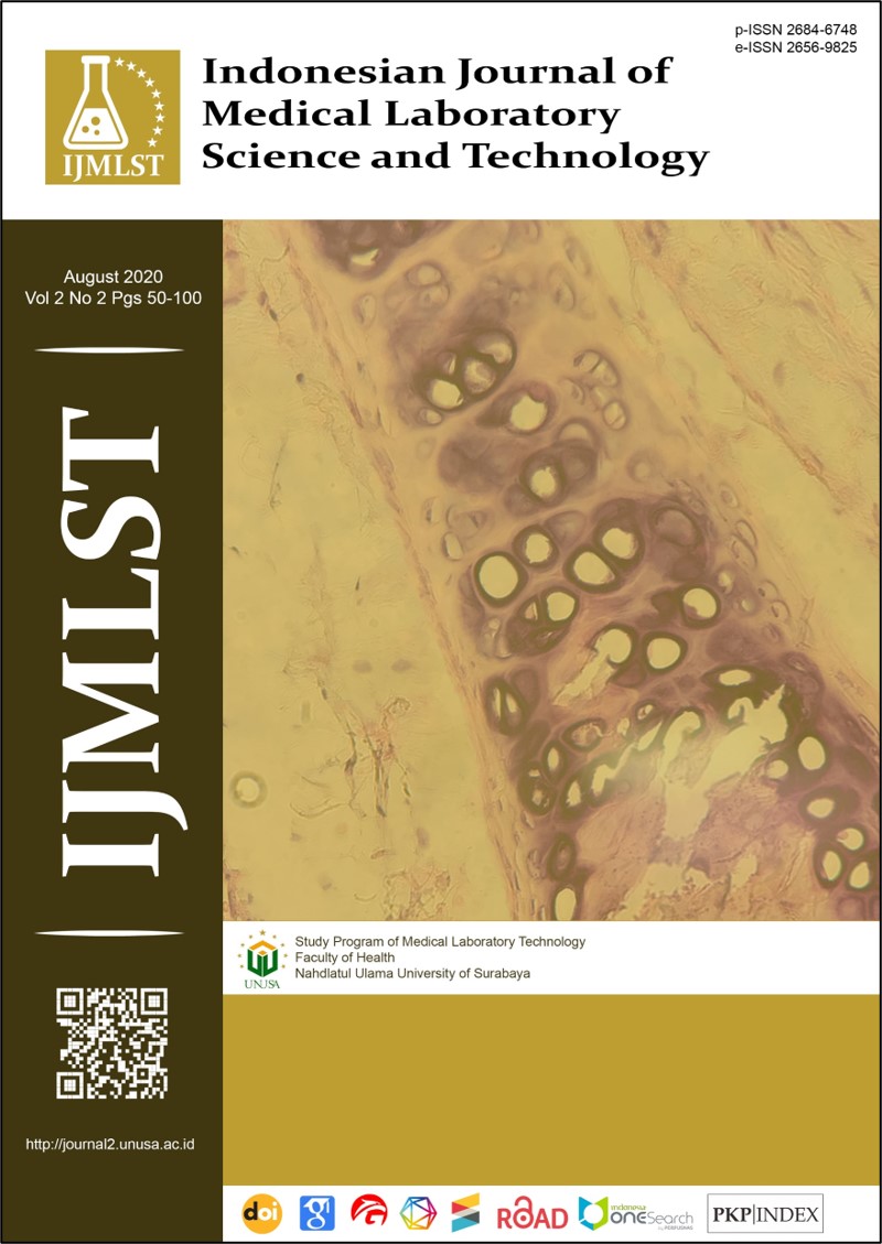
UTILIZATION OF 1% OF METHYLENE BLUE IN STAINING HISTOPATHOLOGICAL PREPARATIONS AT ANATOMIC PATHOLOGY LABORATORY
Main Article Content
Abstract
Tissue staining using hematoxylin-eosin (HE) is a standard method of histopathological staining. The tissue staining is hampered when there is no hematoxylin reagent in laboratory. Therefore, other reagents are needed that can replace the use of hematoxylin. Methylene blue is a basic dyes that interact with cell nuclei which has a negative ionic charge of the tissue. It can be used as an alternative nuclei staining. This study aims to evaluate the use of 1% of methylene blue in cell nuclei staining in histopathological preparations. The research sample were 15 pathology preparations which were randomly selected including breast cancer, cervical cancer and ovarian cancer in the bank of sampel at anatomical pathology laboratory of RSUD Dr. Slamet Garut, Indonesia. The experiment showed that the methylene blue dyes yielded “worth” result (40%) and “poorly” result (60%). Further research can be carried out by modifying the pH of 1% of methylene blue reagent so that it can maximize the staining preparations results as good as those using hematoxylin.
Downloads
Article Details
Copyright (c) 2020 Tri Rahmawati, Yadi Apriyadi, Mamay

This work is licensed under a Creative Commons Attribution-ShareAlike 4.0 International License.
References
Ministry of Health Republic of Indonesia. Cancer Situation. Data and Information Center. 2018
Ankle MR, Kale AD, Charantimath S, Charantimath S. Comparison of staining of mitotic figures by haematoxylin and eosin-and crystal violet stains, in oral epithelial dysplasia and squamous cell carcinoma. Indian J Dent Res. 2007;18:101–105.
Jadhav KB, Ahmed Mujib B R, Gupta N. Crystal violet stain as a selective stain for the assessment of mitotic figures in oral epithelial dysplasia and oral squamous cell carcinoma. Indian J Pathol Microbiol. 2012;55:283-287.
Veuthey, T., Georgina Herrera, G, Dodero, V.I. Dyes and stains: From molecular structure to histological application. Front in Biosci. 2014;19: 91–112.
Titford M, Bowman B. 2012. What May the Future Hold for Histotechnologists? LabMedicine. 2012;43;5–10.
Alturkistani HA, Tashkand FM, Saleh ZMM. Histological Stains: A Literature Review and Case Study. Glob J Health Sci. 2016;8;72–79.
Mescher AL. Histologi Dasar Junqueira : teks & atlas. Ed.12. Jakarta : EGC. 2011.
Suvarna K, Christopher L, Bancroft JD, Theory and Practice of Histological Techniques, 7th edition. Philadhelphia : Elsivier. 2013.
Kadoo P, Dandekar R, Kulkarni M, Mahajan A, Kumawat R, Parate N. Correlation of mitosis obtainned by using 1% crystal violet stain with Ki67LI in histological grades of oral squamous cell carcinoma. J Oral Biol Craniofacial Res. 2017;8;234–240.
Tandon A, Singh NN. Brave, VR Sreedhar G . Image analysis assisted study of mitotic figures in oral epithelial dysplasia and squamous cell carcinoma using differential stains. J Oral Biol Craniofacial Res. 2016;6;18–23.
Soman C, Lingappa A, Mujib, A. Topical Methylene Blue in-vivo Staining as a Predictive Diagnostic and Screening Tool for Oral Dysplastic Changes – A Randomised Case Control Study. Journal of Dental Sccience. 2016;4;118–123.
Kiuchi K. Rapid alkaline methylene blue supravital staining for assessment of anterior segment infections. Clin Ophthalmol. 2016;10;1971–1975.
Bendzinski ECM, Chu K, Johnson JI, Brous M, Copeland K, Bolon B. Optical density-based image analysis method for the evaluation of hematoxylin and eosin staining precision. J Histotechnology. 2020;43;29–37.
Chapman CM. Troubleshooting in the histology laboratory. J Histotechnology. 2019;42;137–149.
Jafari M, Hasanzadeh M. Cell-specific frequency as a new hallmark to early detection of cancer and efficient therapy: Recording of cancer voice as a new horizon. Biomedicine & Pharmacoherapy. 2020;122;109770
Dobrzyńska I, Skrzydlewska E, and Figaszewski Z A. Changes in Electric Properties of Human Breast Cancer Cells. J Membrane Biol. 2013;246; 161–166.


